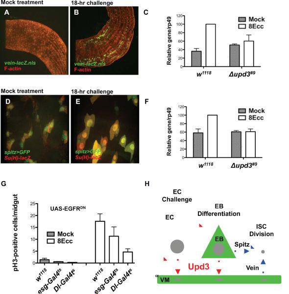
Fig. 7. Role of Upd3 signaling in EGFR-dependent ISC division.
(A–B) Vein is weakly expressed in visceral muscles as visualized by F-actin staining in mock-treated animals and highly expressed in animals infected for 18 hours with Ecc15.
(C) Quantification of vein expression by RT-PCR in mock-treated animals and in animals infected with Ecc15 for 8 hours (8Ecc). For w1118, 8Ecc vs. Mock, P = 0.0006 (***). For Δupd3#9, 8Ecc vs. Mock, P = 0.5585 (ns).
(D and E) Spitz is expressed in EBs as visualized with the Su(H)GBE-lacZ marker in mock-treated animals and in differentiated EBs in animals infected with Ecc15 for 18 hours.
(F) Quantification of spitz expression by RT-PCR in mock-treated animals and in animals infected with Ecc15 for 8 hours (8Ecc). For w1118, 8Ecc vs. Mock, P = 0.0101 (*). For Δupd3#9, 8Ecc vs. Mock, P = 0.8212 (ns).
(G) EGF signaling was conditionally (using tub-GAL80ts) inhibited in ISCs and EBs (esg-GAL4), or ISCs alone (Dl-GAL4) by expressing a dominant-negative form of the EGF receptor (UAS-EGFRDN). For Ecc15 treatment (8Ecc), escg-GAL4 vs. w1118, P = 0.0414 (*) and Dl-GAL4 vs. w1118, P = 0.0012 (**).
(H) Model of intestinal homeostasis. Erwinia infection activates upd3 expression in mature enterocytes (EC) and in differentiating enteroblasts (EB) (Upd3 production and producing cells are depicted in red). The basal secretion of the Upd3 cytokine activates JAK/STAT signaling in EBs and in visceral muscles (VM) (JAK/STAT signaling depicted in green). Activation of JAK/STAT signaling in EBs and VMs leads to EGF ligand production that stimulates the EGFR-dependent division of ISCs (EGF signaling depicted in blue).

