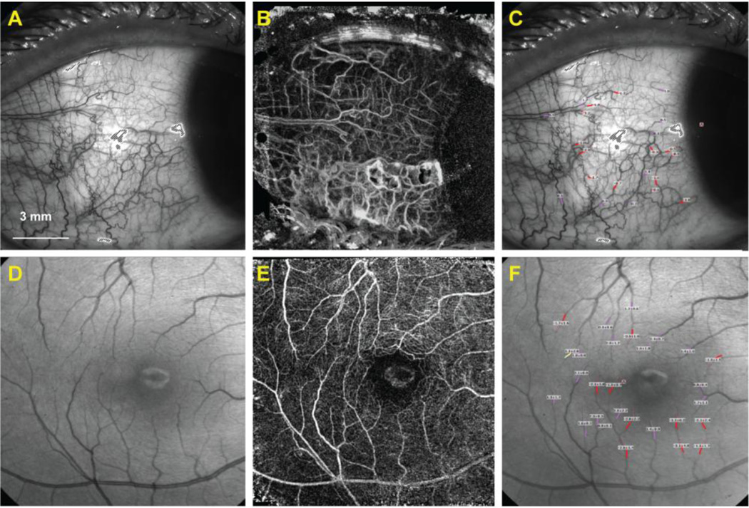
Figure 1. Human retinal and conjunctival microvascular assessed with a retinal function imager (RFI).
The bulbar conjunctiva (A) of a healthy subject was imaged, and measurements of the conjunctival capillary perfusion (B) and blood flow velocity (unit: mm/s) (C) were obtained. Similarly, the fovea of another healthy subject (D) was imaged, and measurements of the capillary perfusion (E) and blood flow velocity expressed in mean ± SD (unit: mm/s) (F) were obtained. The avascular zone was evident in the fovea. Note that negative values (red) indicate blood flow moving away from the heart and that the vessels are arteries. Positive values (pink) represent the veins.

