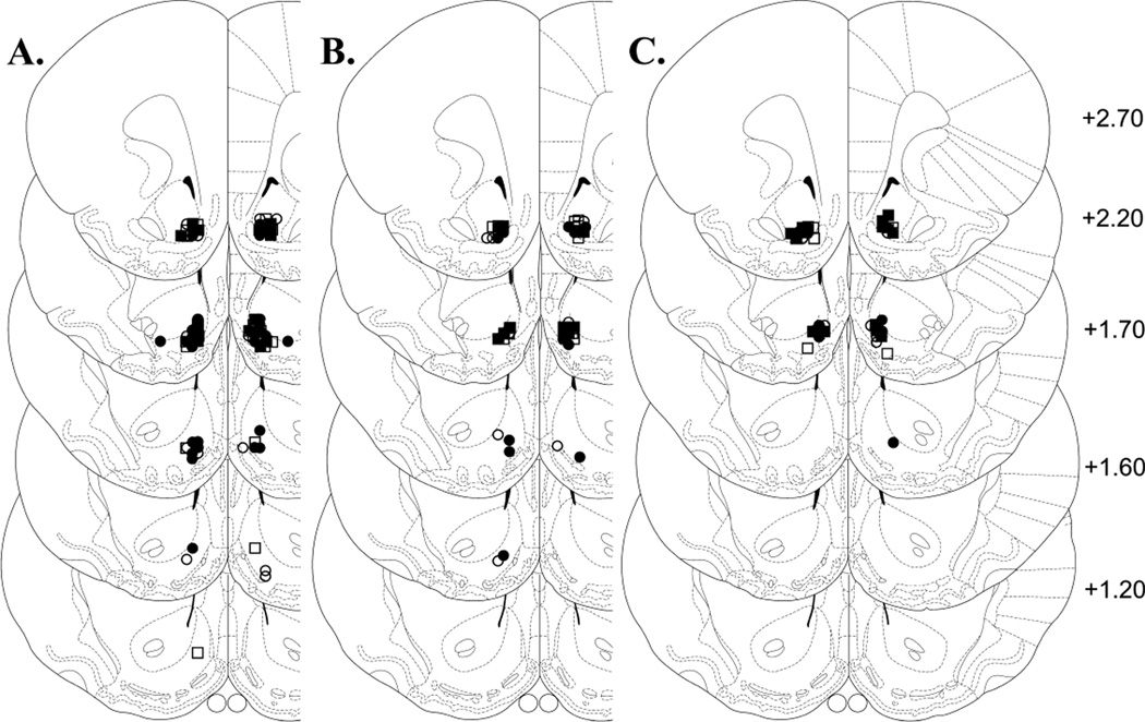
Fig. 3.
Location of microinjection cannula tips in the NAcc shell for all rats included in the data analyses. Line drawings of coronal sections (adapted from Paxinos and Watson, 1997) show the location of the microinjection cannula tips in the NAcc shell for rats tested with no inhibitor (A), with SCH23390 (B) or with Rp-cAMPS (C). Numbers to the right indicate mm from bregma. Symbols denote group affiliation: filled circles, αCaMKII-AMPA; open circles, control-AMPA; filled squares, αCaMKII-saline; open ssquares, control-saline.

