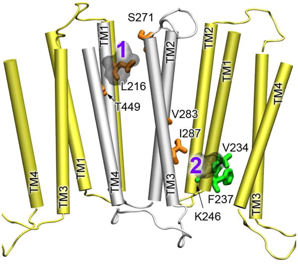
Fig. 4.
The ketamine-binding sites in the α4β2 nAChR. The TM domains of α4 and 2 are colored in yellow and silver, respectively. The residues of α4 and β2 showing changes in chemical shift upon halothane binding are highlighted in green and orange sticks, respectively. The docked ketamine molecules are numbered and shown in light gray.

