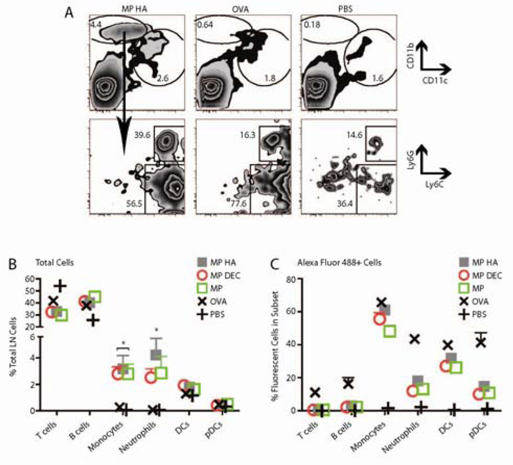
Fig 4. Fluorescent particles recruit neutrophils and monocytes to the draining LN.
10µg microparticles encapsulating OVA Alexa Fluor 488 were injected s.c. in both hind footpads. Mice were sacrificed after 1 day, and popliteal LNs were taken. Cells were stained for CD11c, CD11b, B220, TCRβ, CD4, CD8, Ly6G and Ly6C. A. Graphs shown were previously gated on live cells (FSC vs. SSC). Top Row. All microparticle groups caused a large recruitment of CD11bhi CD11clo cells that was not observed in OVA- or PBS-injected mice. Shown are representative FACS plots from MP HA−, OVA−, and PBS-injected mice. Bottom Row. CD11bhi CD11clo cells were both neutrophils and monocytes. B–C. Data was combined from two experiments, three mice each. B) The percentage of monocytes and neutrophils was increased in microparticle- versus OVA-injected popliteal LNs. C) The efficiency of particle or OVA uptake for each cell subset.

