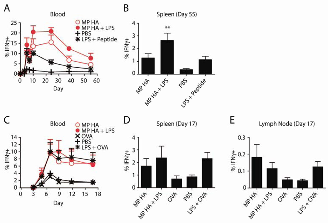
Fig 5. VSV-OVA challenge following vaccinations produces increased IFNγ+ CD8 T cells.
BMDCs were incubated overnight with 0.85µg/mL MP HA, 0.85µg/mL MP HA + 0.42µg/mL LPS, 0.42µg/mL LPS + 5µM SIINFEKL (OT-I peptide), or PBS control. 250,000 of these BMDCs were given to mice i.v. 7 days post-BMDC injection, mice were challenged with 105 PFU VSV-OVA i.v. and A) the T cell response was tracked via blood on days 0, 3, 5, 7, 10, 24, 38, and 55 post-challenge. Lymphocytes from blood were purified, restimulated with OT-I peptide, and stained for CD4, CD8, IFNγ, CD44, B220, Ter119, and CD11b. Antigen-specific cells were detected by IFNγ+CD44+ staining. Data shown is previously gated on CD8+ T cells. B) At day 55, mice were sacrificed, spleens were taken, and antigen-specific cells were detected as in blood. One-way ANOVA was done with Bonferroni post-hoc testing. C–E. Direct immunogenicity. Mice were given 10µg MP HA, 10µg MP HA + 10µg LPS, 10µg LPS + 10µg EndoGrade OVA, 10µg EndoGrade OVA, or PBS s.c. in both hind footpads. At day 45, mice were challenged with 105 PFU VSV-OVA i.v., C) and subsequently bled at days 3, 5, 7, 9, 12, and 17. Lymphocytes from blood were purified, restimulated with OT-I peptide and stained for CD4, CD8, IFNγ, CD44, B220, and CD11b. Antigen-specific cells were detected by IFNγ+CD44+ staining. Data shown is previously gated on CD8+ T cells. D–E) At day 17, mice were sacrificed, spleens and LNs were taken, and antigen-specific cells were detected as in blood.

