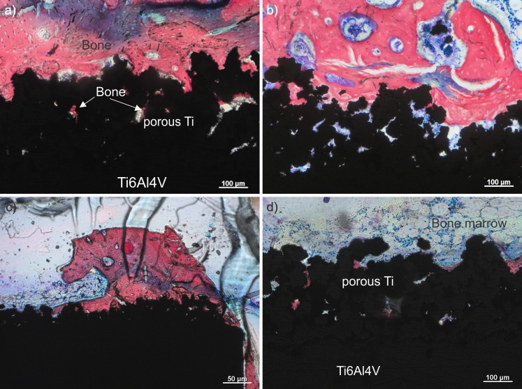
Fig. 5.
Characteristic histological sections of the samples with bioactive glass (BAG) (a) and without BAG (b). c Sample with BAG at lower magnification and part of the implant in the bone marrow (d). Mineralized bone is stained red. Osteoblasts and bone marrow cells in b are stained blue. The blue region in the upper part of the image (a) is an artifact caused during the staining with Stevenel’s blue

