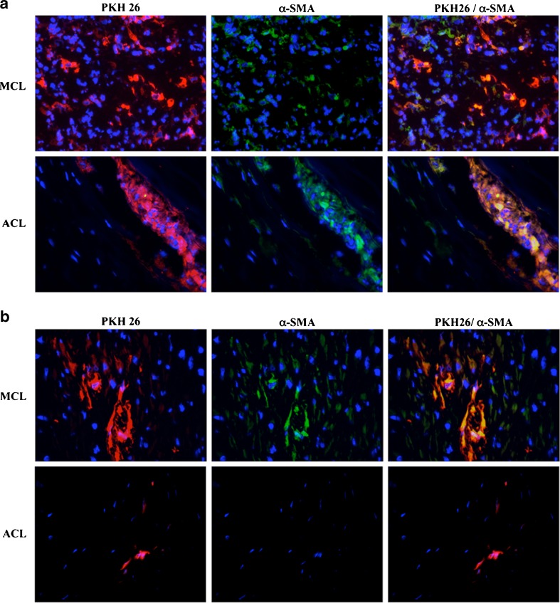
Fig. 2.
PKH-26+ cells maintained their myofibroblasts phenotype in vivo up to 7 days after transplantation in rabbit ligament. Culture-derived myofibroblasts labelled with PKH-26 (red) were transplanted in damaged [medial collateral ligaments (MCL)] or intact [anterior cruciate ligament (ACL)] ligaments, and biopsies were performed at 1 (a) or 7 (b) days after cell injection. Tissue sections were analysed using immunofluorescence for α-smooth-muscle actin (α-SMA) (green) and 4’-6’-diamidino-2-phenylindole (DAPI) (blue). The sections shown are representative of five analysed grafts

