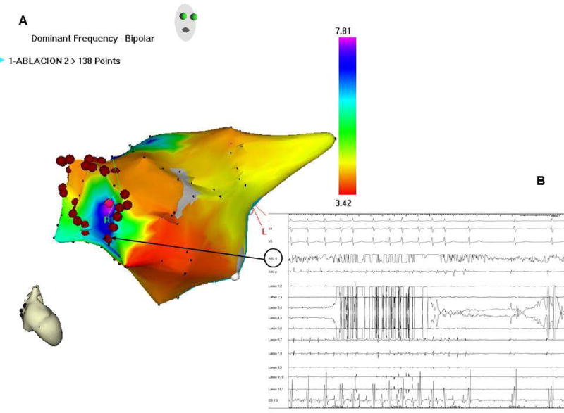
Figure 2.
A: Real-time atrial DF map (right anterior view; CARTO system) in a paroxysmal AF patient. Purple, high DF site on right superior PV antrum. Red dots, circumferential ablation line. B: Surface ECG leads and intracardiac lasso catheter electrograms within RSPV; CS and ablation catheter (black arrow) during radiofrequency delivery, with sinus rhythm conversion, prior to isolation of the RSPV, confirming the reentrant nature of AF, incompatible with the multiple wavelet theory. CS: coronary sinus; DF: dominant frequency; ECG: electrocardiogram; PV: pulmonary vein. (Unpublished).

