Fig. 2.

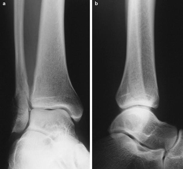
AP and lateral radiographic images of a SE-2 fracture, consisting of a spiral or oblique fibula fracture at the level of the syndesmosis

AP and lateral radiographic images of a SE-2 fracture, consisting of a spiral or oblique fibula fracture at the level of the syndesmosis