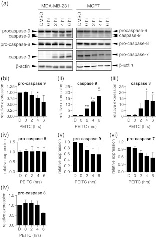
Fig. 3.
Caspase expression in PEITC-treated cells. MDA-MB-231 or MCF7 cells were treated with PEITC (20 μM) for the indicated times or DMSO for 6 h as a control. Expression of caspase 9, caspase 8, caspase 3 (MDA-MB-231 cells) and caspase 7 (MCF7 cells) was analysed by immunoblotting. β-actin was analysed as a loading control. a Representative immunoblots. The positions of migration of pro- and cleaved forms of caspases are indicated. b Quantitation. Data shown are means ± SD derived from two independent experiments for pro-caspase 9, caspase 9, caspase 3 and pro-caspase 8 in MDA-MB-231 cells (i–iv, respectively) and pro-caspase 9, pro-caspase 7 and pro-caspase 8 in MCF7 cells (v–vii, respectively). Expression values were normalized to that of β-actin. Statistically significant differences between DMSO and PEITC treated cells are indicated (*p < 0.05; **p < 0.005). Active caspase 9 was not detected in MCF7 cells

