Fig. 2.
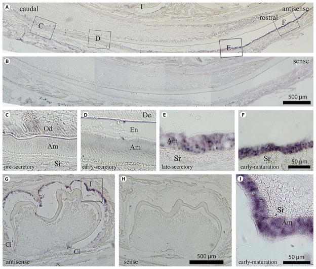
In situ hybridization analysis of NCKX4 in the developing mandibular dentition of a 7-day-old mouse. Mouse incisors (A–F) and molars (G–I) in sagittal section using the antisense riboprobe (A, C–F, G, I) versus the sense (negative control) riboprobe (B, H). No hybridization signal is evident in the negative control sections. C–F, I, are enlarged regions identified in A and G. Ameloblast cell populations are presecretory (C), early-secretory (D), late-secretory (E) and early-maturation (F, I). The highest level of NCKX4 mRNA is seen during late-secretory and maturation-stage amelogenesis as evidenced in the incisors (E and F compared to C and D) and in the molar (I). Am = Ameloblasts; Cl = cervical loop; De = dentin; En = enamel; Od = odontoblasts; Sr = stellate reticulum. A, B, G, H Scale bars: 500 μm. C–F, I Scale bars: 50 μm.

