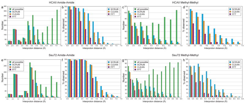
Figure 5.
Removal of aliasing artifacts promotes peak identification in the 4-D TS NOESY spectra for HCAII (top) and Ssu72 (bottom). NOE crosspeaks with intensities over four times the noise level in spectra processed with either the FT, CLEAN, or SCRUB were matched to their corresponding interproton distances, and unique distances are plotted as histograms in (a, c, e, g). Each interproton distance was counted as observed if either corresponding NOE (Hi → Hj or Hj → Hi) surpassed the noise threshold. “All expected” refers to the total number of interproton distances calculated from a reference crystal structure (PDB code 2ILI for HCAII and PDB code 3P9Y for Ssu72). The histograms in (b, d, f, h) show the percent of observed distances relative to all expected. Short interproton distances that were not identified in the SCRUB-processed methyl-methyl spectra are the result of signal degeneracy. For example, the four unidentified distances under 3.5 Å (out of 53 total) for HCAII correspond to intraresidue methyl-methyl NOE crosspeaks for residues V109, V121, L143, and L223 that overlap with diagonal signals. The small number of very long distances (>8.0 Å) observed in the methyl-methyl spectrum of Ssu72 likely result from incorrect rotamer assignments in the reference crystal structure due to the insufficient resolution of the model (2.1 Å) for precisely defining ILV sidechain orientations. For example, the two longest distances—9.31 Å between I173 δ1 and L176 δ2 and 8.55 Å between V43 γ2 and L45 δ2—are substantially shorter in a second crystal structure (PDB code 3FDF, 3.2 Å resolution)—6.99 Å and 7.87 Å, respectively—as the result of different rotamer selections for residues L45 and I173.

