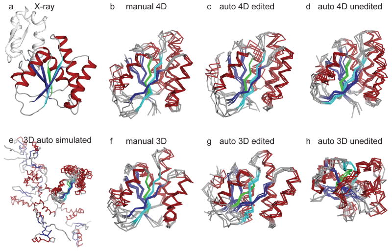
Figure 7.
CYANA structure calculations for Ssu72 using manually assigned and manually edited peaks (b, f), auto-assigned and manually edited peaks (c, g), and auto-assigned and unedited peaks (d, h). In (b–d), peak lists are from SCRUB-processed 4-D TS spectra. Green and cyan coloring highlights the characteristic β-propeller twist of the central β-sheet that is lost in the ensemble produced by auto-assigned and manually edited 3-D peak lists (compare (c) and (g)) In (f–h), peak lists are from conventional 3-D TS spectra. In (e), calculations are based on simulated 3-D peak lists derived from 4-D spectra. The reference crystal structure (PDB code 3FDF) is shown in (a). The substrate-binding subdomain of Ssu72, which is not constrained to the main phosphatase domain by the TS NOESY data, is omitted from (b–h), and is aligned separately in Figure S2. The ensemble in (e) is aligned over the largest segment of converged structure.

