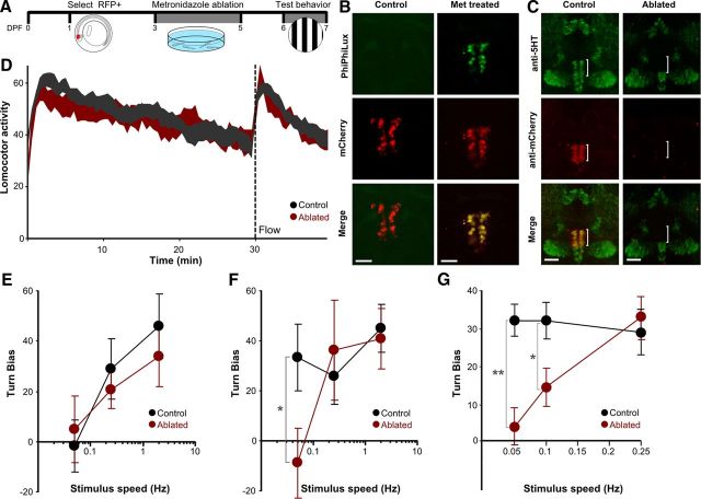
Figure 5.
Ablation of the DR blocks enhanced visual sensitivity during arousal. A, Timeline for experiments using metronidazole to ablate the DR in Tg(tph2:nfsB-mCherry)y226 fish. B, Detection of activated caspase-3 during ablation using PhiPhiLux G1D2 (green) reveals ongoing apoptosis selectively in DR mCherry-positive cells in metronidazole-treated (right) but not vehicle-treated (left) larvae. Scale bar, 25 μm. C, Confocal stacks in control (left) and metronidazole-ablated (right) fish. Immunohistochemistry against mCherry (red) and serotonin (green). Scale bar, 50 μm. D, Locomotor activity of control (gray) and ablated (red) fish (n = 9 × 20). A 60 s flow stimulus was applied at 30 min (dotted line). E, F, Turn bias in response to optomotor stimuli presented 5 min before (E) and 3 min after (F) the flow stimulus in control (black) and ablated (red) larvae (n = 14 × 20). *p < 0.05. G, Turn bias of the OMR during transfer arousal in control (black) and DR-ablated (red) larvae (n = 9 × 25). *p < 0.05, **p < 0.001. Graphs show mean and SEM.

