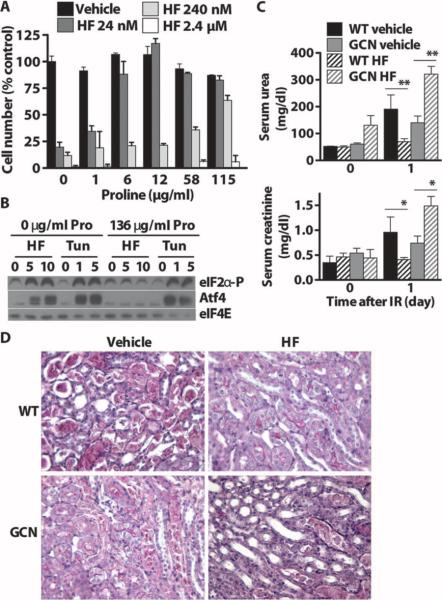
Fig. 6.
Block of HF activity by excess proline in vitro and requirementfor Gcn2 in vivo. (A) Cytostatic/cytotoxic effects of increasing concentrations of HF added to proliferating MEF cultures and blockade by addition of excess proline to the medium as measured by the MTT assay and expressed as a percentage of cell number in the vehicle-treated group with no additional proline. (B) Phospho-eIF2α and Atf4 immunoblots of extracts prepared from MEFs treated with HF (0, 5, or 10 nM) or tunicamycin (Tun) (0, 1, or 5 μg/ml) for 3 hours in complete medium with or without proline (Pro) supplementation. eIF4E was used as a loading control. (C) Protection by HF against renal IR required Gcn2. Serum urea (top) and creatinine (bottom) before (day 0) and 1 day after 25 min of bilateral renal IR. Error bars indicate SEM. Asterisks indicate the significance of the difference between the indicated groups by Student's t test for effect of diet within the same genotype. *P < 0.05; **P < 0.01. (D) Representative images of PAS-stained kidney sections 1 day after reperfusion showing the corticomedullary junction, where most tubular damage occurs.

