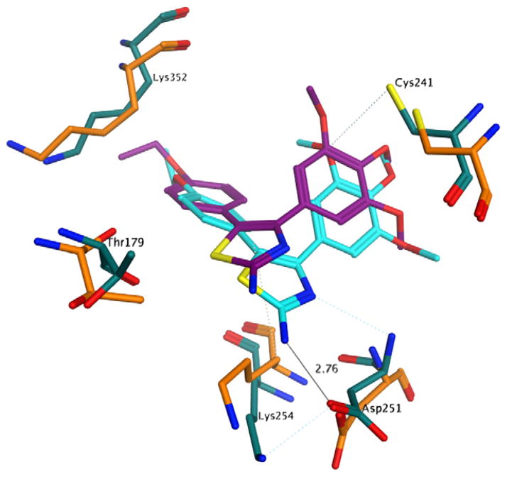
Figure 1.
Proposed binding pose of 2e to tubulin. Before the molecular dynamics: carbon atoms of 2e in purple, carbon atoms of tubulin residues in orange; after the molecular dynamics: carbon atoms of 2e in cyan, carbon atoms of tubulin residues in turquoise. Other atoms indicated as follows: red, oxygen; blue, nitrogen; yellow, sulphur; Hydrogen atoms are not shown.

