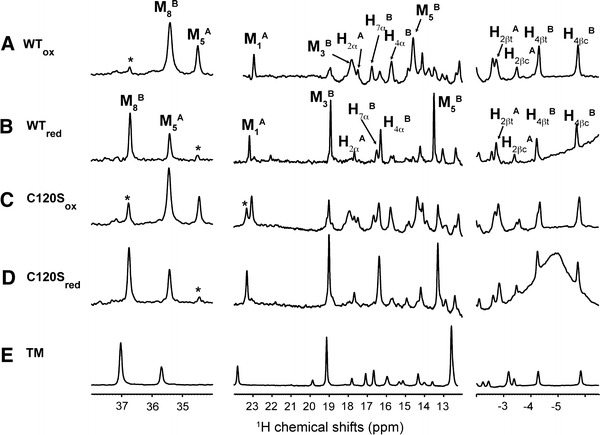
Fig. 2.

One-dimensional 1H NMR spectra of the Ngb protein. Spectra A and B were collected on the wild-type (WT) Ngb protein without dithiothreitol treatment (WT ox) or after dithiothreitol treatment (WT red), respectively. Spectra C and D are the spectra of oxidized and reduced C120S Ngb. Spectrum E is the 1D spectrum of C46G/C55S/C120S triple-mutant (TM) Ngb. All NMR spectra were collected at 298 K in 20 mM tris(hydroxymethyl)aminomethane–HCl buffer (pH 7.4) at 100 μM protein concentration using a 600-MHz NMR spectrometer. The assigned heme atoms are labeled for WT Ngb in the WTox and WTred states. Peaks labeled with a star correspond to incomplete reduction or oxidation of the protein
