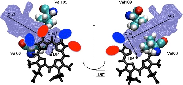
Fig. 6.

Close-up schematic view of the heme region of Ngb. The heme skeleton is represented as black sticks. The mechanically sensitive Val68 (E11) and Val109 (G8) residues are represented as cyan spheres and the Xe2/Xe4 cavities and the distal pocket (DP) are indicated as an ice blue surface. In this scheme, the red and blue ovals indicate the volumes occupied by the vinyl groups of the A and B heme conformers, respectively. Consequently, the red ovals and blue ovals indicate the volumes occupied by methyl groups of the B and A heme conformers, respectively. The view on the right was obtained from a 180° rotation of the same scheme around the vertical axis
