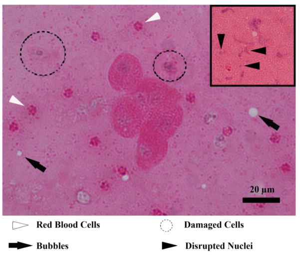
Figure 11.
H&E stain of the collected fountain projectiles. In the centre of the image, a cell cluster consisting of six whole cells is present. In addition, there are red blood cells (white arrowheads), damaged or dying cells (dotted circles), and vapour bubbles (black arrows). The insert shows smeared and fragmented nuclei (black arrowheads).

