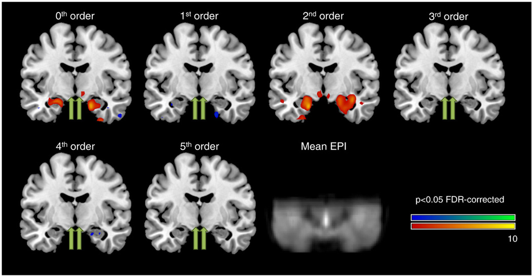
Fig. 1.
Coronal plane (MNIy=−8 mm) overlaid with areas of significant parametric modulations for 0th to 5th order (p<0.05 FDR-corrected). In accordance with the hypothesis, significant second-order modulation was found in left and right hypothalamus (yellow arrows) as well as bilateral amygdala. For comparison, hypothalamic second-order modulation maxima are also indicated for all other modulation orders (green arrows). For details please refer to Table 1. In addition, the group-averaged EPI is also shown to indicate brain coverage and to demonstrate the high quality of fMRI data in hypothalamus/amygdala regions.

