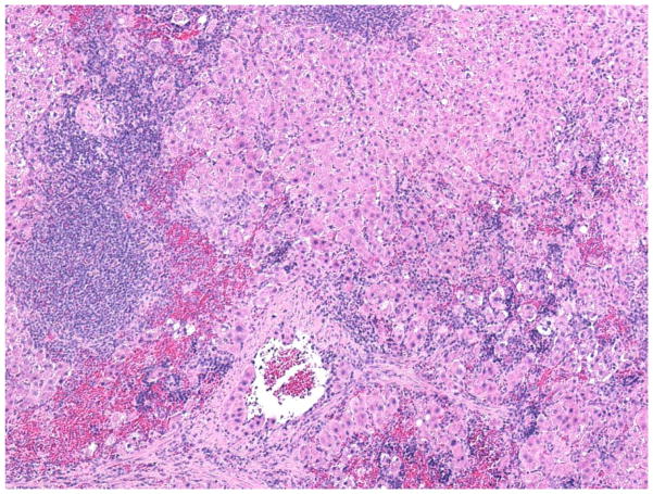
Fig. 1.
Morphological appearance of intrasplenically transplanted hepatocytes. Light microscopic appearance of intrasplenic hepatocytes 45 weeks after transplantation (×200). Spleens were fixed in 10% buffered formalin and stained with hematoxylinand eosin. Large masses of hepatocytes within the red pulp occupy -70% of the figure (from left to right).

