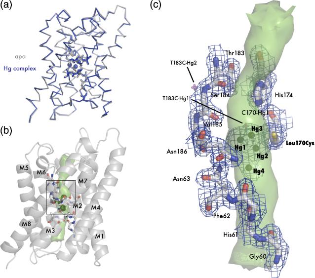
Figure 4.
Crystal structure of apo L170C and mercury bound L170C. (A) Main chain overlay of the apo (gray) and Hg-complex (blue) with an RMSD of 0.27 Å. Bound mercury atoms are displayed as spheres with a van der Waals radius of 1.10 Å. (B) Cartoon representation of L170C. Transmembrane helices are labeled M1-M8 and the interior surface of the channel is drawn as a green surface. The black square denotes the area of interest depicted in panel C. (C) Structure of the blocked channel. Amino acids classically involved with water binding in AQPs are shown as sticks and with 2Fo-Fc - electron density mapped contoured at 1.2 σ drawn in blue. Mercury are shown as spheres. Superposition of mercury atoms from the T183C structure are shown as magenta crosses. In this orientation it can be seen that all three mercury atoms sterically block the pore (green surface).

