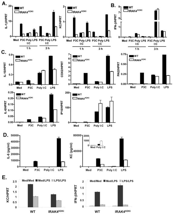
Figure 1. Cytokine gene expression in WT versus IRAK4KDKI macrophages.
WT and IRAK4KDKI macrophages were treated with medium only (Med) or stimulated with Pam3Cys (P3C; 1 μg/ml), Poly I:C (40 μg/ml), or LPS (100 ng/ml) for 1 or 3 h and analyzed for MyD88-dependent (A) and MyD88-independent (B) gene expression by qPCR. (C) Macrophages were treated as in (A) and (B) for 3 h and additional gene expression was analyzed by qPCR. (D) Macrophages were treated as in (A–C) for 6 h and supernatants were collected for cytokine measurement. (E) Macrophages isolated from WT and IRAK4KDKI mice were treated with medium only (Med/Med and Med/LPS) or LPS (100 ng/ml; LPS/LPS) for 18 h, washed 3 times then re-stimulated with medium (Med/Med) or LPS (100 ng/ml; Med/LPS and LPS/LPS) for 5 h. MyD88-dependent (KC) and MyD88-independent (IFN-β) gene expression was analyzed by qPC R. All samples were normalized to HPRT. Data are representative of at least 3 separate experiments.

