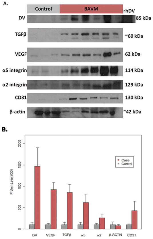
Fig. 1.

Protein levels of DV, VEGF, TGFβ, α5, α2, and CD31 are increased in human BAVM compared to control (epilepsy) tissue. (A) Representative western blots for each protein investigated. Note that recombinant human DV (rhDV) was used as a positive control for DV blots. (B) Bar graph displaying the mean amounts of DV (p < .001), VEGF (p < .001), TGFβ (p < .001), α5 (p = .001), α2 (p = .006), and CD31 (p = .02) were increased in BAVM patients compared to controls, while β-actin was not (p = .649). These relationships remained the same after adjusting for CD31 and β-actin in the ANCOVA models for DV (p = .04), VEGF (p < .001), TGFβ (p = .008), α5 (p = .05), and α2 (p = .03).
