Figure 4.
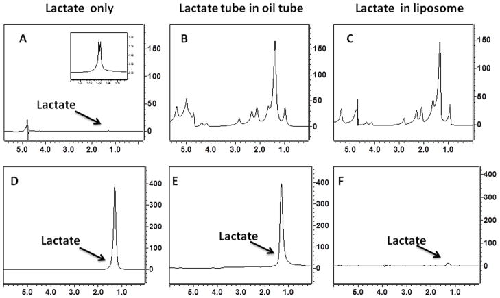
Water suppressed pulse acquire (A, B, C) and HadSelMQC (D, E, F) spectra of 10 mM lactate phantoms. (A) Phantom 1: lactate in aqueous solution, (B) Phantom 2: an aqueous solution of lactate inserted into a tube with vegetable oil, (C) Phantom 3: lactate in a liposomal suspension. Large lipid signals in phantoms 2 and 3 (panel B and C) completely obscure the lactate signal (A). The HadSelMQC sequence filtered out the lipids signals and revealed the underlying lactate resonance (panels E and D). A difference in signal intensity in top and bottom panels is due to differences in receiver gain: 2 and 60 dB for spectrum A, B, C and D, E, F respectively. No differences in signals intensity were detected in experiments in phantoms containing only lactate (D) or in lactate in the presence of oil but not in direct physical contact with it (E). The liposome provides a large surface area for integration between lactate and lipid head groups. Such an interaction leads to reduction of lactate T2 and reduction of NMR visibility of lactate (panel F).

