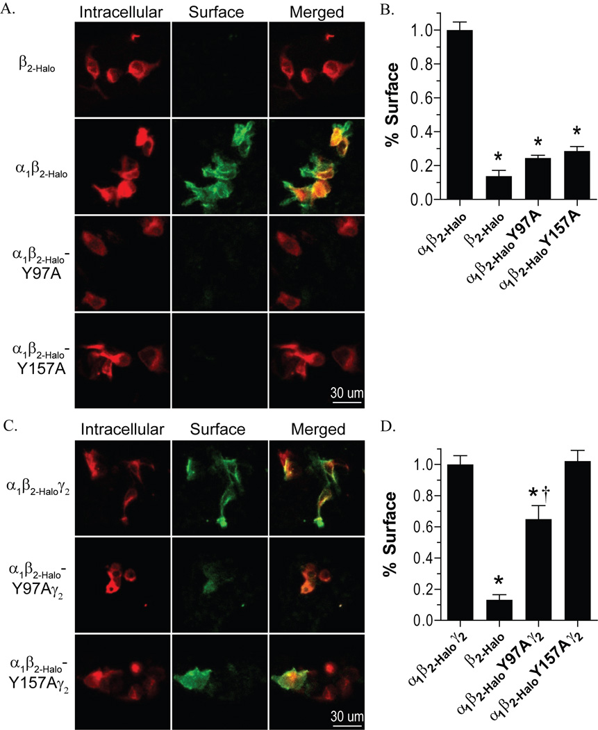
Figure 3.
Surface expression of receptors containing mutant β2 subunits. A and B) Expression of Halo-tagged subunits was determined by labeling with a cell-impermeable Alexafluor 488 ligand (green), followed by labeling with a cell-permeable TMR ligand (red), and images were collected by confocal microscopy. C and D) The percent of surface expression was quantified by measuring the mean intensity of the AlexaFluor 488 label relative to the total of AlexaFluor 488 label plus TMR label mean intensity of individual cells. The percent surface expression is normalized to the wild-type α1β2-Halo receptor (C) or α1β2-Haloγ2 receptor (D). * Denotes significant difference when compared to the wild-type receptor and † denotes a significant difference when compared to the negative control (β2-Halo only), Student’s t-test (p < 0.05).

