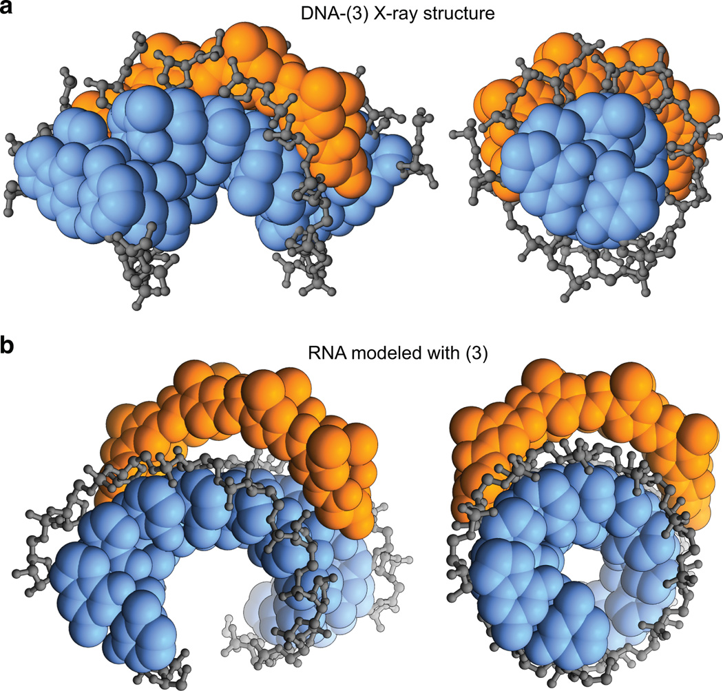
Figure 3.
Structural basis for selective dsDNA versus dsRNA binding. a) Crystal structure of DNA-polyamide 3 complex (PDB: 3OMJ) showing shape complementary and favorable hydrophobic interactions with the sugar-phosphate backbone (gray). Orange, polyamide; blue, aromatic DNA bases. b) Coordinates of cyclic polyamide 3 docked in the minor groove of the putative binding site on a model of ideal A-form dsRNA.

