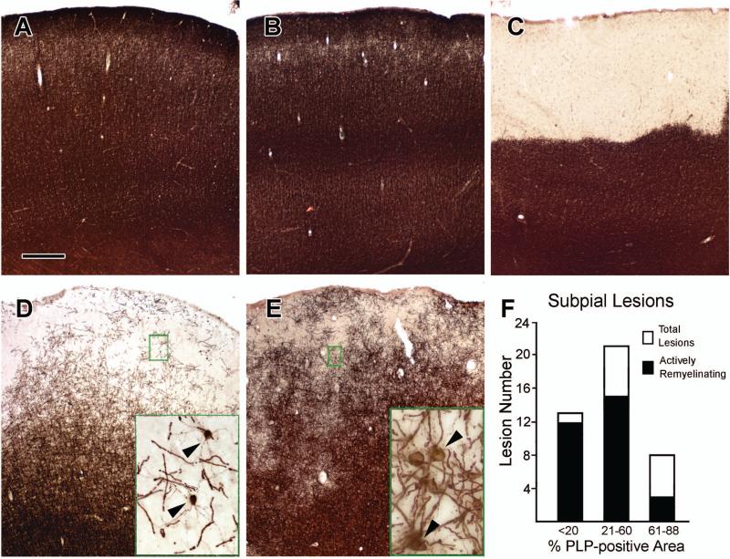
Figure 1. Remyelination in Subpial Cortical Lesions.
Brains from control patients without neurological disease have dense PLP immunoreactivity in all cerebral cortical layers (Panel A). Many regions of cortex in patients with MS show a similar pattern in PLP immunoreactivity (Panel B). A subpial demyelinated lesion with complete loss of PLP immunoreactivity is shown in Panel C. Many subpial lesions are not completely demyelinated, but contain PLP-positive myelin internodes (Panels D and E). Frequently, these areas contain actively-myelinating oligodendrocytes with PLP-positive cell bodies and processes extending to myelin internodes (Panels D and E, arrowheads in insets). Actively-myelinating oligodendrocytes are most frequent in lesions where PLP-positive myelin occupied 60% or less of the lesion area (Panel F). The scale bar represents 400 μm for Panels A-E.

