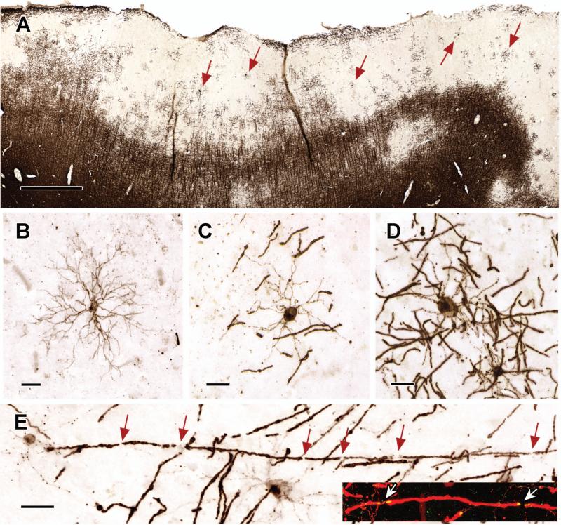
Figure 2. Active Remyelination in Subpial Cortical Lesions.
Subpial cortical lesions contain oligodendrocytes in various stages of differentiation (Panel A, arrows). Premyelinating oligodendrocytes with PLP-positive cell bodies and multiple processes are present (Panel B), but actively-myelinating oligodendrocytes with PLP-positive cell bodies and processes extending to multiple myelin internodes are more prevalent (Panels C and D). Myelin-internodal lengths are relatively short (Panels C and D), which supports the interpretation that these oligodendrocytes are actively remyelinating axons. Individual axons are ensheathed by multiple-short myelin internodes (Panel E, inset, arrows indicate nodes of Ranvier). Remyelinated fibers also show molecular maturation (Panel E, inset) as demonstrated by the appropriate distribution of the paranodal protein, Caspr (yellow in Panel E, inset; red in inset is PLP staining). The scale bar in Panel A represents 1 mm; the scale bars in Panels B - E represent 20 μm.

