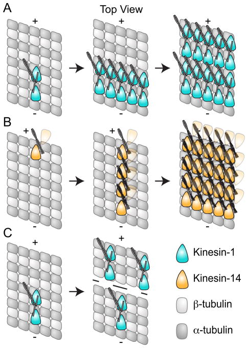
Figure 6. Cooperative binding to the Microtubule.
A. Kinesin-1 (cyan) dimers exhibit cooperative binding to the microtubule, and the presence of 16 nm layer lines in cryo-EM suggests this proceeds in an lateral manner (Vilfan et al., 2001). Kinesin-14 dimers (orange) also display cooperative binding; however in contrast to Kinesin-1, this appears to proceed along the protofilament (Wendt et al., 2002; Cope et al., 2010). The second head in the Kinesin-14 dimer is depicted as partially transparent since cooperative binding is only observed when one head of the dimer is bound; for Kar3Vik1, binding of both heads in the ADP state appeared to abolish cooperative binding (Rank et al., 2012). C. Kinesin-1 dimers also exhibit long range cooperative binding where binding of one dimer biases additional dimers to bind further towards the plus-end of the microtubule (Muto et al., 2005).

