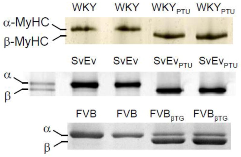
Figure 1.

Characteristics of cardiac myofibrillar proteins. Each lane represents one heart, and examples from two hearts are shown for each population. Gel electrophoresis stained with Coomassie-Blue demonstrated ~100% α-MyHC expression in hyperthyroid Wistar Kyoto (WKY) rats and normal 129/SvEv wildtype mice (SvEv), while ~100% β-MyHC was expressed in the PTU-fed WKY rats and SvEv mice. Transgenic mice expressing ~70% β-MyHC (FVBβTG) were compared against non-transgenic FVB controls expressing 100% α-MyHC to test for any potential complications of PTU diet.
