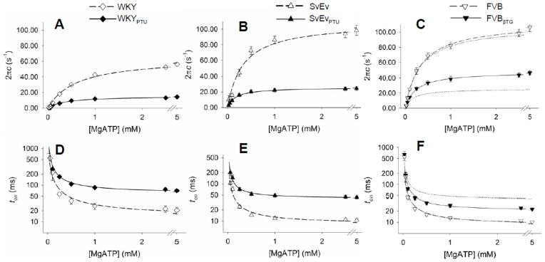
Figure 4.
Sensitivity of myosin crossbridge detachment rate, 2πc, and time-on, ton, to MgATP. A–C. At all MgATP concentrations greater than 0.1 mM, myosin detachment rate was significantly higher for α-MyHC compared to β-MyHC in rats (panel A) and mice (panels B and C). Detachment rates for 70% β-MyHC in the FVBβTG mouse were lower than 100% α-MyHC, but not as low as 100% β-MyHC (Panel C, where dotted lines reflect curves from Panel B). D–F. At all MgATP concentrations greater than 0.1 mM, mean myosin crossbridge ton was shorter in the α-MyHC compared to β-MyHC. For the same isoform myosin crossbridge ton was shorter in mice compared to rats.

