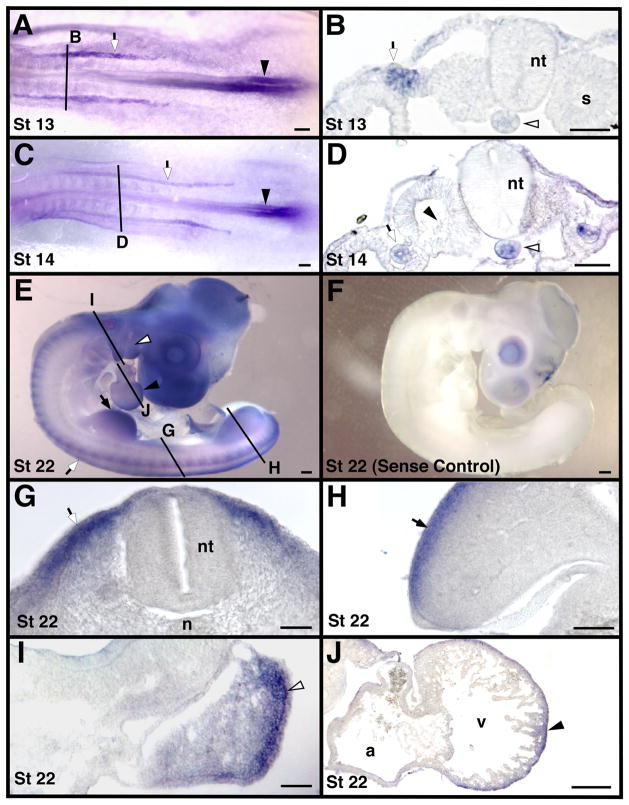
Figure 4.
Localization of MT2-MMP mRNA in whole mount and transverse sections of stage 13 – 22 chicken embryos. Solid black lines indicate approximate cross-section level shown in corresponding labeled images. Panel F is an embryo hybridized with a sense probe, all other panels are embryos hybridized with an antisense probe for MT2-MMP. Shown are stages 13 in panels A and B, 14 in panels C and D, and 22 in panels E–J. Caudal notochord expression continues until approximately stage 14 (white arrowheads in B and D) as does neural fold expression (black arrowheads in A and C). The intermediate mesoderm (white arrows in A–D) also expressed MT2-MMP. MT2-MMP was expressed in the dermamyotome (white arrows in E and G) and in the limb (black arrows in E and H). The pharyngeal arches (white arrowheads in E and I) and the heart (black arrowheads in E and J) also expressed MT2-MMP. Note the lack of expression in the sclerotome (black arrowhead in D). Scale bar = 50 μm in B, D, G, H, I and J and 100 μm in A, C and E. Abbreviations: a, atrium; n, notochord; nt, neural tube; s, somite; and v, ventricle.

