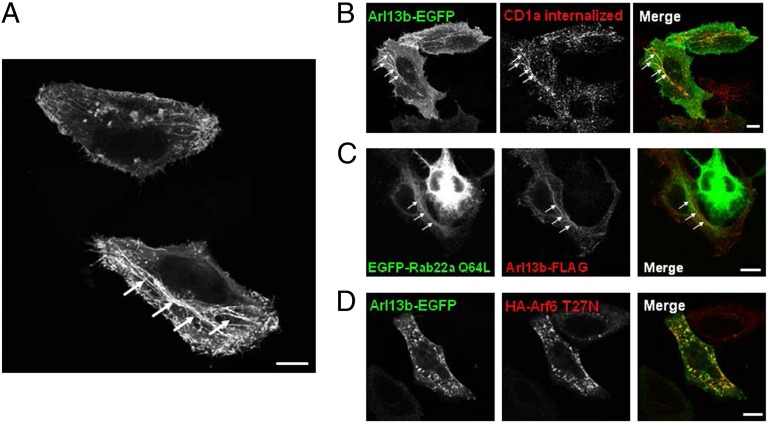
Fig. 3.
Arl13b-EGFP colocalizes with CD1a, Rab22a, and Arf6 in HeLa cells. (A) HeLa cells were transiently transfected with Arl13b-EGFP and analyzed by confocal microscopy. Arrows indicate Arl13b-decorated tubular structures. (B) HeLa cells were cotransfected with CD1a and Arl13b-EGFP and incubated with anti-CD1a mAb for 30 min on ice. Cells were then washed and incubated for 30 min at 37 °C. After the surface-bound Ab was stripped in a brief acidic wash, cells were fixed, permeabilized, and labeled with Alexa Fluor 546-conjugated anti-mouse secondary Ab to detect anti-CD1a (red). (C) HeLa cells were transiently cotransfected with EGFP-Rab22a Q64L and Arl13b-FLAG, fixed, permeabilized, and stained with Cy3-conjugated mouse monoclonal anti-FLAG Ab (red). (D) HeLa cells were transiently cotransfected with Arl13b-EGFP and HA-Arf6 T27N, fixed, permeabilized, and stained with anti-HA tag polyclonal Ab followed by Alexa Fluor 546-conjugated anti-rabbit secondary Ab (red). (Scale bars: 10 μm.)

