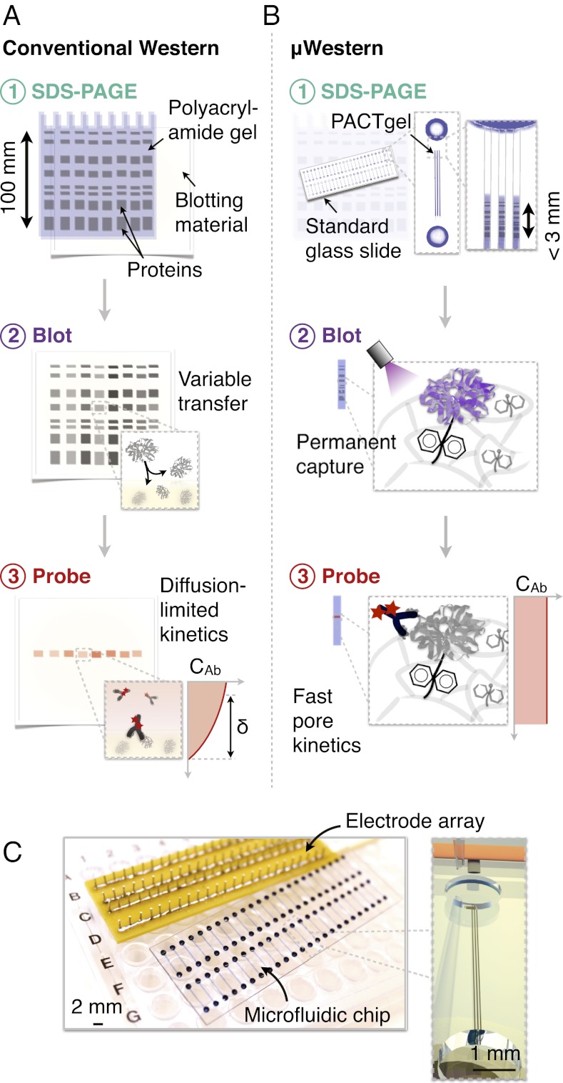
Fig. 1.
Single-microchannel μWestern assay design enables high device density formats. Aspects of scale, reagent use, blotting efficiency, and probe-binding kinetics are illustrated by comparative schematics for the conventional (A) and μWestern (B) assays (δ indicates a diffusion boundary layer thickness). The microfluidic workflow is comprised of: (i) analyte stacking and SDS-PAGE within the PACTgel matrix; (ii) band capture (“blotting”) onto the benzophenone-decorated PACTgel in response to UV light (as opposed to transfer to a separate sheet of hydrophobic material in conventional Western blotting); (iii) removal of SDS by brief electrophoretic washing and electrophoretic introduction of fluorescently labeled primary and (optionally) secondary detection antibodies specific to the target. Finally, excess probe is electrophoretically driven out of each device and peak intensities determined by fluorescence micrograph analysis. (C) Modular interfacing of standard microscope slide-sized chips with a scalable electrode array accommodating 48 blots per chip in triplicate (144 microchannels).

