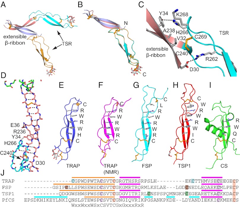
Fig. 3.
The extensible β-ribbon and TSR domains. Views of the extensible β ribbon and TSR domain of the two chains in the P. vivax open-TRAP structure after superposition on the VWA domain (A) or TSR domain (B). (C) The interface between the extensible β ribbon and TSR domain. Key sidechains are shown as sticks and hydrogen bonds as dashes. (D) The backbone of extensible β ribbon with key sidechains and disulfides as sticks, and hydrogen bonds as dashes. (E–I) TSR domains. Structures in identical orientations are from our vivax TRAP crystal structure (E), an isolated falciparum TRAP TSR domain NMR structure (F) (4), and F-spondin (G) (48), thrombospondin-1 (H) (16), and CS (I) (2). Layer residues in TSR domains are shown as sticks and labeled. (J) Sequence alignment of representative TSR domains. Cysteines that disulfide link are colored identically. Colored overlines mark strand 1 (orange), β-strands (magenta), and helices (cyan).

