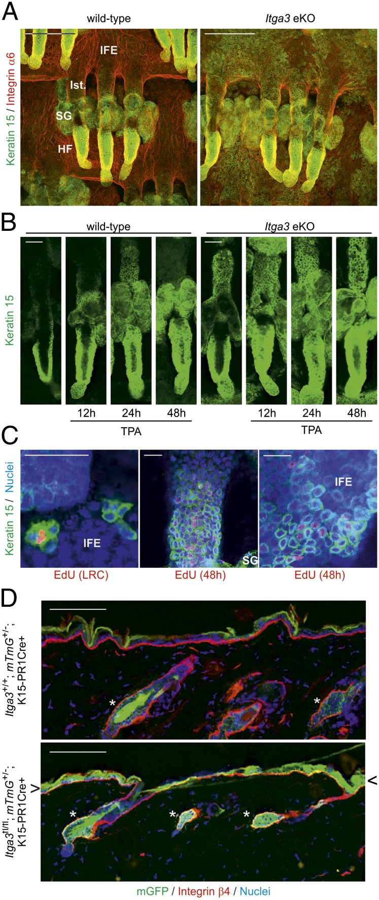
Fig. 4.
Keratin 15–positive cells from the HF bulge exit their niche in the absence of Itga3. (A) HF and IFE of whole-mounted Itga3-null tails are Krt15+, whereas this cell population is confined to the HF bulge in WT littermates (Ist., isthmus). (Scale bar, 100 μm.) (B) Keratin 15 stainings of whole-mounted tail HFs at indicated time points after single TPA doses. Twenty-four hours after TPA, Krt15+ cells have migrated out of the bulge into the isthmus of WT HFs. In Itga3 eKO mice, basal efflux of Krt15+ cells increases further on TPA stimulation. (Scale bar, 50 μm.) (C) (Left) EdU+ LRC in IFE is Krt15+. (Center and Right) Forty-eight hours after pulse, EdU+ Krt15+ cells are present in the isthmus and IFE. (Scale bar, 50 μm.) (D) HF bulge cells from the back skin of Itga3fl/fl; mTmG+/−; K1-15-CrePR1+ mice leave their compartment (asterisk) and migrate into the IFE (arrowheads). They remain in the bulge region (asterisk) in WT littermates. (Scale bar, 100 μm.)

