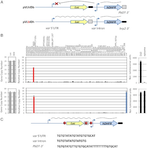
Fig. 1.
A DNA element found in the PbDT 3′ UTR disrupts the intron’s ability to silence a var promoter. Stably transfected parasite lines carried constructs mimicking var gene structure with luciferase as a reporter gene for var promoter expression and hdhfr as a positive selectable marker expressed by the var intron. (A) (Upper) Schematic of the pVLhIDb episome that contains a var promoter paired with a var intron. This var promoter is properly regulated and silent by default. (Lower) Schematic of the pVLbIDh episome, which is identical to pVLhIDb except for the replacement of the 3′ UTR that terminates luciferase. This replacement resulted in activation of the var promoter, which no longer is recognized by the mechanism that controls mutually exclusive expression. (B) Results of pVLhIDb (Upper) and pVLbIDh (Lower) in the DC-J parasite line. Quantification of the ratio between var promoter and intron by qRT-PCR (Left). Steady-state mRNA levels of each individual var gene measured by qRT-PCR are presented as copy number relative to the housekeeping genes arginyl-tRNA synthetase (PFL0900c) (Center) and the levels of luciferase expression (Right). The D10 parasite line (22) constitutively expressing luciferase from an endogenous var promoter was used as a positive control. All values presented are the average of at least two biological replicates. Error bars represent SE. (C) A similar TG-rich DNA element is found within the PbDT 3′ UTR, var promoter, and var intron. In the schematic of the construct, the TG-rich element is shown as a red circle, the PbDT 3′ UTR is boxed, and the hrp2 3′ UTR is shown as a bold black line.

