Fig. 2.
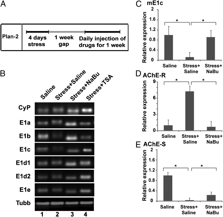
Differential regulation of AChE exons after FSS and drug treatment. (A) Schematic representation of the course of the experiment. After the FSS protocol had been followed for 4 d, the mice were returned to their original cages for 1 wk and then were injected with NaBu, TSA, or saline daily for 1 wk and were killed 2 wk thereafter (n = 4). (B) Semiquantitative RT-PCR analysis of the AChE 5′ transcripts (mE1a–mE1e) in the hippocampus of control or stressed mice treated with saline or an HDACi (NaBu or TSA). Lane 1, saline; lane 2, stress + saline; lane 3, stress + NaBu; lane 4, stress + TSA. NaBu completely restored the down-regulation of mE1b and mE1c exons at the 5′ end. TSA, on the other hand, showed variable effects. (C–E) Real time PCR of mE1c (C), AChE-R (D), and AChE-S (E) in the hippocampus of control mice, stressed mice, and stressed mice treated with either saline or NaBu. At the 3' end, similar restoration was found in AChE-R mRNA levels, while AChE-S mRNA levels were partially restored with NaBu treatment. *P < 0.05, 2-tailed u test.

