Fig. 2.
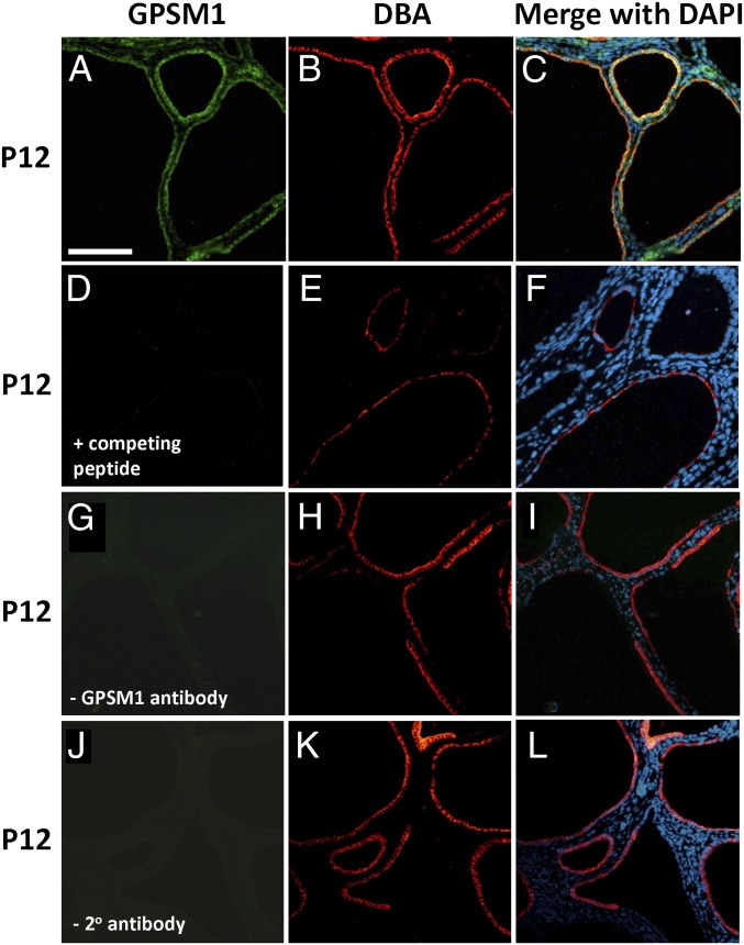
GPSM1 localization in cystic ADPKD mouse kidneys by immunofluorescent histochemistry. Serial sections were made from kidneys harvested at postnatal day 12 from cystic Gpsm1+/+;Pkd1V/V mice. Kidney sections were stained with GPSM1 (green color in A) and a collecting duct lectin (DBA; red color in B, E, H, and K). Nuclei were stained with DAPI (blue color in C, F, I, and L). Negative controls for GPSM1 staining were sections incubated with competing peptide (D), no primary GPSM1 antibody (G), and no secondary antibody (J). Magnification 40×. (Scale bar: 0.1 mm for all images.)

