Fig. 5.
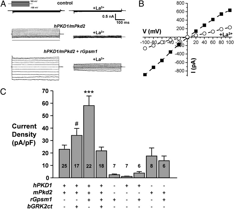
GPSM1 enhances PC1/PC2 activity. (A) Typical macroscopic currents before and after La3+ under voltage-clamp conditions from nontransfected (control) CHO cells or transfected with human PKD1 (2 μg) and mouse Pkd2 (1 μg) either alone or coexpressed with rat Gpsm1 (1 μg). Currents were elicited by test pulses with 20-mV steps from 100 to −100 mV from a holding potential of 0 mV. Voltage pulse protocol is shown in the inset. (B) Representative macroscopic I/V relation before (closed squares) and after (open circles) La3+ from the experiment presented in A. (C) Summary graph of the La3+-sensitive current at –100 mV for voltage-clamped CHO cells expressing PC1 and PC2 in the absence and presence of wild-type rat GPSM1 (rGpsm1) or bovine GRK2ct (bGRK2ct). The number of observations in each group is shown in each bar. ***P < 0.001 versus all groups; #P < 0.05: significant difference between hPKD1/mPkd2 + rGpsm1 and hPKD1/mPkd2 + bGRK2ct.

