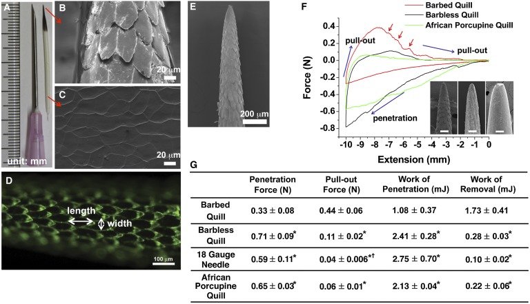
Fig. 1.
Geometrical features of the North American porcupine quill and analysis of the penetration and removal forces with muscle tissue. (A) North American porcupine quill. (B and C) FE-SEM images showing the microstructure of the quill tip and base, respectively. (D) Fluorescence image enables visual delineation of the geometry of single barbs. (E) FE-SEM image showing the microstructure of the tip of the porcupine quill. (F) Representative force versus extension plots show puncture, penetration, and removal of barbed, barbless, and African porcupine quills from muscle tissue (see Fig. S3A for experimental set-up). (Inset) Micron-level topography of the three quills. (Scale bars: 100 μm.) Red arrows indicate resistance as the barbed quill is removed from the tissue (not observed for others). (G) Summary of experimental values obtained from penetration/removal of barbed quill, barbless quill, 18 gauge needle, and African porcupine quill (mean ± SD, n = 5). Each mean is compared with every other mean using one-way ANOVA with Tukey’s Honestly Significant Difference post hoc analysis to correct multiple comparisons at 95% confidence interval by using GraphPad Prism 6 (*P < 0.05, compared with barbed quill; †P < 0.05, compared with barbless quill).

