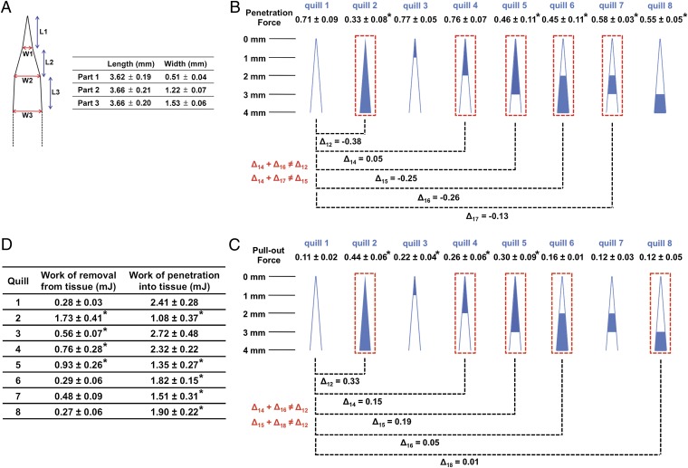
Fig. 4.
Barbs within a 4-mm barbed region at the apex of the quill work independently to minimize penetration force and cooperatively to maximize pull-out from tissue. (A) Dimensional analysis of the porcupine quill through length scale measurements of natural quills (mean ± SD, n = 5). In terms of curvature, there are three transition points. L and W indicate length and width, respectively. (B, C) Penetration and pull-out forces were obtained with the prepared quills via sanding to isolate the contribution of barbs within different regions (see Fig. S5) (mean ± SD, n = 5). The penetrating depth for all experiments was 10 mm (see Fig. S3A for the experimental set-up). Cartoons depict quills prepared with specific lengths of barbs obtained through ablation with sand paper. The blue color indicates the barbed region and the white color indicates the barbless region. The penetration and pull-out forces of the prepared quills are compared with those of the barbless quill (quill 1). The difference in force is defined as Δij (Δij = penetration (or pull-out) force of quill j–penetration (or pull-out) force of quill i)). (D) Summarized work of penetration and work of removal obtained through penetration–retraction tests with muscle tissue (mean ± SD, n = 5). All are compared with quill 1 (*P < 0.05, one-way ANOVA Fisher’s Least Significant Difference post hoc analysis at 95% confidence interval by using GraphPad Prism 6).

