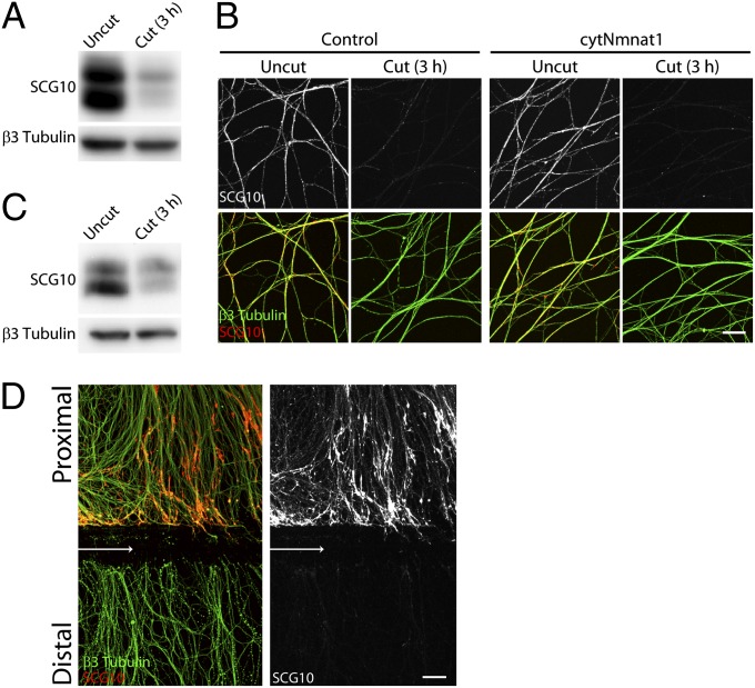
Fig. 1.
SCG10 loss is an early marker of axonal injury. (A) Immunoblot analysis of endogenous SCG10 in cultured DRG axons with or without axotomy. The SCG10 level is decreased dramatically in the distal axons collected 3 h after axotomy compared with the level in the uncut control axons. Immunoblot against neuron-specific β3 tubulin confirms comparable amounts of protein loaded. (B) Axonal SCG10 is examined by immunostaining 3 h after axotomy in the DRG cultures infected with control or cytNmnat1-expressing lentivirus. The SCG10 level is decreased by axotomy to a similar extent in the distal axons of cytNmnat1-expressing neurons and in the control culture. β3 tubulin (green) labels microtubules in axons. (Scale bar, 50 µm.) (C) Three hours after sciatic nerve transection in adult mice, SCG10 levels were assayed by immunoblot in a control nerve and in the nerve segment distal to the axotomy. The SCG10 level is decreased significantly by axotomy. β3 tubulin is shown as a loading control. (D) SCG10 is lost selectively in the distal axons as shown by immunolabeling against SCG10 and β3 tubulin in the DRG cultures 3 h after axotomy. Arrows indicate the site of axotomy. SCG10 protein is accumulated in the proximal segment. (Scale bar, 50 µm.)

