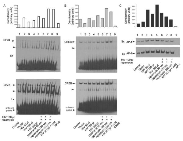
Figure 5. HIV induced activation of NF-kB, CREB and AP-1 transcription factors.
EMSA was carried out using the nuclear extracts prepared from CIDHPs following treatment with HIV pseudotyped virus for 5 h either in the presence or absence of rapamycin (100 nM). The densitometric scanned data for NF-kB, CREB, and AP-1 are expressed on an arbitrary scale. In each set, the last lane is a repetition of conditions using nuclear extracts following 200 μl HIV virus treatment of podocytes, however the reaction mix pre-incubated with 10 ng (cold) unbiotinylated probe, before adding the biotinylated probe, demonstrate the specificity of respective labeled DNA oligomer. For clarity, the biodyne membranes were exposed to the X-ray film either for longer (5-10 min) or for shorter (1-2 minutes) time periods. The top shows the picture of the autoradiogram of the shorter exposure time (Sx) and the bottom shows the longer exposure time (Lx). Densitometry data of DNA-protein complexes was quantified and shown as a histogram.

