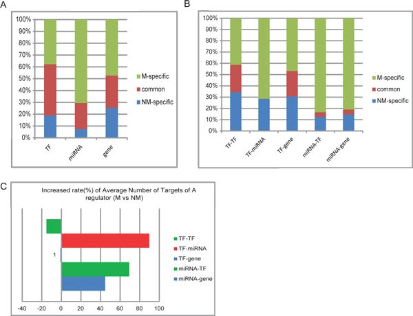
Figure 2.
Comparison of global regulatory patterns between the HCC non-metastatic and metastatic networks. (A)(B). Comparison of node- and edge- distributions between the HCC metastatic and non-metastatic networks. Nodes or edges were divided into three categories: only in non-metastatic network(NM-specific), only in metastatic network(M-specific), and common in both networks. (C). Increased rate of average number of targets of a regulator in the metastatic network versus the non-metastatic. For each TF- or miRNA- relations as a whole, average number of targets was calculated in each network, and then the increased rate of the average number of targets in the metastatic network versus the non-metastatic one was represented in barplot. The color of the bar represents the type of targets, TF in green, miRNA in red, and gene in blue.

