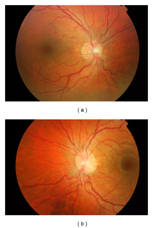
Figure 2.

Retinal photographs of the right (a) and left (b) eyes 1 week after the initial visual symptoms. In the right optic disc, the limits are blurred more prominently in the superior and inferior poles. In the left eye optic disc, contour is almost normal.
