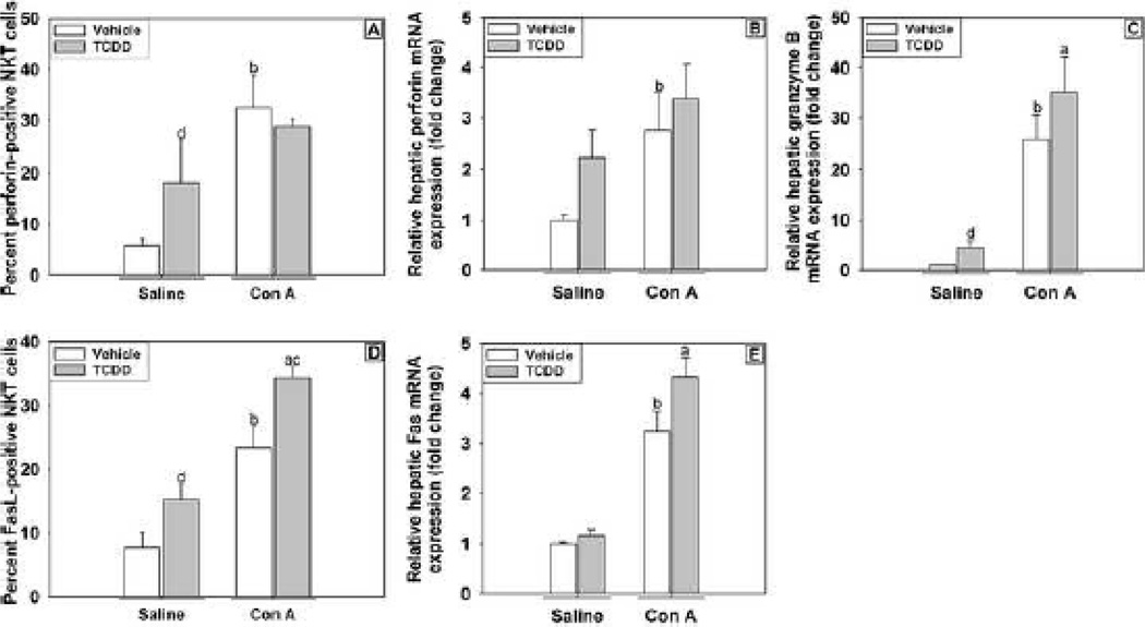
Figure 9. Expression of cytolytic markers on hepatic NKT cells and hepatic expression of perforin, granzyme B and fas in mice treated with TCDD/Con A.
Mice were treated as described in the legend to Figure 2, and hepatic NKT cells (NK1.1+, CD3ε+) were isolated from mice and stained for markers of cytolytic activity. (A) NKT cells isolated 3 h after Con A or Saline administrations were stained for intracellular perforin. (D) NKT cells isolated 4 h following Con A or Saline administration were stained for expression of Fas Ligand (CD95). Liver samples were collected 6 h after Saline or Con A treatment, and hepatic perforin (B), granzyme B (C) and Fas (E) mRNA expression was determined by RT-PCR. a p < 0.05 TCDD/Con A versus TCDD/Saline. b p < 0.05 Vehicle/Con A versus Vehicle/Saline. c p < 0.05 TCDD/Con A versus Vehicle/Con A. d p < 0.05 TCDD/Saline versus Vehicle/Saline. Data represent the mean ± SE of independent replicates 2 separate experiments. For each treatment group: Vehicle/Saline n=3–6, TCDD/Saline n=3–6, Vehicle/Con A n=3–6, TCDD/Con A n=3–6.

