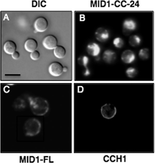
Fig 2.

Loss of the modulatory region of Mid1 alters the localization pattern of Mid1 in C. neoformans. (A) Mid1-CC-24–DsRed fusion protein was constructed by tagging the Mid1-CC-24 truncated mutant with DsRed (28) and visualized by DIC and confocal microscopy. (A) A DIC image of cryptococci expressing the Mid1-CC-24–DsRed fusion protein revealed bright, spherical cells of C. neoformans. (B and C) Interestingly, Mid1-CC-24–DsRed appeared to be mislocalized. This was in stark contrast to the full-length Mid1 (Mid1-FL) dsRed fusion protein, which is predominately localized to the cell surface of C. neoformans, consistent with its plasma membrane distribution as previously reported (2). (D) Immunofluorescence (Texas Red) of Cch1 revealed a surface distribution in C. neoformans similar to that of Mid1, confirming that, like Cch1, Mid1 is primarily localized to the plasma membrane in cells of C. neoformans. A primary peptide antibody to the C terminus of Cch1 and a Texas Red-conjugated secondary antibody were used to visualize Cch1 as reported previously (2, 4). The specificity of the primary peptide antibody to Cch1 was confirmed in previous publications (2, 4). Scale bars represent 10 μM.
